Back Angiografie Afrikaans تصوير الأوعية Arabic Angioqrafiya Azerbaijani آنژیوقرافی AZB Ангіяграфія Byelorussian Angiografia Catalan Angiografie Czech Angiografi Danish Angiografie German Angiografio Esperanto
This article needs more reliable medical references for verification or relies too heavily on primary sources. (November 2021) |  |
| Angiography | |
|---|---|
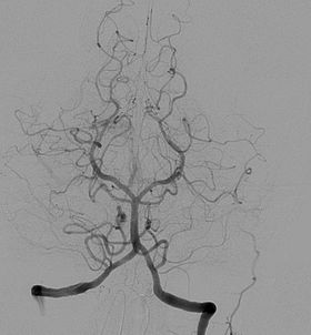
 Angiogram of the brain showing a transverse projection of the vertebro basilar and posterior cerebral circulation. | |
| ICD-9-CM | 88.40-88.68 |
| MeSH | D000792 |
| OPS-301 code | 3–60 |
Angiography or arteriography is a medical imaging technique used to visualize the inside, or lumen, of blood vessels and organs of the body, with particular interest in the arteries, veins, and the heart chambers. Modern angiography is performed by injecting a radio-opaque contrast agent into the blood vessel and imaging using X-ray based techniques such as fluoroscopy.
The word itself comes from the Greek words ἀγγεῖον angeion 'vessel' and γράφειν graphein 'to write, record'. The film or image of the blood vessels is called an angiograph, or more commonly an angiogram. Though the word can describe both an arteriogram and a venogram, in everyday usage the terms angiogram and arteriogram are often used synonymously, whereas the term venogram is used more precisely.[1]
The term angiography has been applied to radionuclide angiography and newer vascular imaging techniques such as CO2 angiography, CT angiography and MR angiography.[2] The term isotope angiography has also been used, although this more correctly is referred to as isotope perfusion scanning.
- ^ G. Timothy Johnson, M.D. (1986-01-23). "Arteriograms, Venograms Are Angiogram Territory". Chicago Tribune. Retrieved 12 September 2011.
- ^ Martin, Elizabeth (2015). "Angiography". Concise Medical Dictionary (9th ed.). Oxford: Oxford University Press. doi:10.1093/acref/9780199687817.001.0001. ISBN 9780199687817.