Back غرفة العين الأمامية Arabic অক্ষিগোলকের সম্মুখ প্রকোষ্ঠ Bengali/Bangla Prednja očna komora BS Cambra anterior Catalan Πρόσθιος θάλαμος του βολβού Greek Cámara anterior Spanish اتاقک قدامی چشم Persian Etukammio Finnish Chambre antérieure de l'œil French Cuaisín tosaigh Irish
| Anterior chamber of eyeball | |
|---|---|
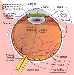
 Anterior part of human eye, with anterior chamber at right. | |
 Schematic diagram of the human eye. | |
| Details | |
| Identifiers | |
| Latin | camera anterior bulbi oculi |
| Acronym(s) | AC |
| MeSH | D000867 |
| TA98 | A15.2.06.003 |
| TA2 | 6792 |
| FMA | 58078 |
| Anatomical terminology | |
The anterior chamber (AC) is the aqueous humor-filled space inside the eye between the iris and the cornea's innermost surface, the endothelium.[1] Hyphema, anterior uveitis and glaucoma are three main pathologies in this area. In hyphema, blood fills the anterior chamber as a result of a hemorrhage, most commonly after a blunt eye injury. Anterior uveitis is an inflammatory process affecting the iris and ciliary body, with resulting inflammatory signs in the anterior chamber. In glaucoma, blockage of the trabecular meshwork prevents the normal outflow of aqueous humour, resulting in increased intraocular pressure, progressive damage to the optic nerve head, and eventually blindness.
The depth of the anterior chamber of the eye varies between 1.5 and 4.0 mm, averaging 3.0 mm. It tends to become shallower at older age and in eyes with hypermetropia (far sightedness). As depth decreases below 2.5 mm, the risk for angle closure glaucoma increases.
- ^ Cassin, B.; Solomon, S. (1990). Dictionary of eye terminology. Gainesville, Fla: Triad Pub. Co. ISBN 978-0-937404-33-1.