Back Wortelvlies (tandheelkunde) Afrikaans ألياف الأنسجة الداعمة Arabic Wurzelhaut German Ligamento periodontal Spanish لیگامان دور دندانی Persian Ligament alvéolo-dentaire French ליגמנט פריודונטלי HE Gyökérhártya Hungarian Legamento parodontale Italian 歯根膜 Japanese
| Periodontal ligament | |
|---|---|
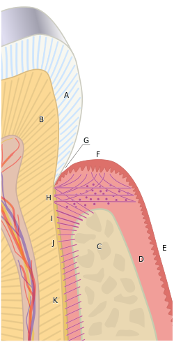
 The tissues of the periodontium combine to form an active, dynamic group of tissues. The alveolar bone (C) is surrounded for the most part by the subepithelial connective tissue of the gingiva, which in turn is covered by the various characteristic gingival epithelia. The cementum overlaying the tooth root is attached to the adjacent cortical surface of the alveolar bone by the alveolar crest (I), horizontal (J) and oblique (K) fibers of the periodontal ligament. | |
| Details | |
| Precursor | Dental follicle |
| Identifiers | |
| Latin | fibra periodontalis |
| Acronym(s) | PDL |
| MeSH | D010513 |
| TA2 | 1611 |
| FMA | 56665 |
| Anatomical terminology | |
The periodontal ligament, commonly abbreviated as the PDL, are a group of specialized connective tissue fibers that essentially attach a tooth to the alveolar bone within which they sit.[1] It inserts into root cementum on one side and onto alveolar bone on the other.
- ^ Wolf HF, Rateitschak KH (2005). Periodontology. Thieme. pp. 12–. ISBN 978-0-86577-902-0. Retrieved June 21, 2011.