Back مهاد Arabic Talamus Azerbaijani تالاموس AZB Таламус Byelorussian Зрочны груд BE-X-OLD Таламус Bulgarian কক্ষ (আন্তরমস্তিষ্ক) Bengali/Bangla Talamus BS Tàlem Catalan تالامەس CKB
| Thalamus | |
|---|---|
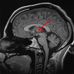
 Thalamus marked (MRI cross-section) | |
 Visual depiction of basic thalamus | |
| Details | |
| Part of | Diencephalon |
| Parts | See List of thalamic nuclei |
| Artery | Posterior cerebral artery and branches |
| Identifiers | |
| Latin | thalamus dorsalis |
| MeSH | D013788 |
| NeuroNames | 300 |
| NeuroLex ID | birnlex_954 |
| TA98 | A14.1.08.101 A14.1.08.601 |
| TA2 | 5678 |
| TE | E5.14.3.4.2.1.8 |
| FMA | 62007 |
| Anatomical terms of neuroanatomy | |
The thalamus (pl.: thalami; from Greek θάλαμος, "chamber") is a large mass of gray matter on the lateral walls of the third ventricle forming the dorsal part of the diencephalon (a division of the forebrain). Nerve fibers project out of the thalamus to the cerebral cortex in all directions, known as the thalamocortical radiations, allowing hub-like exchanges of information. It has several functions, such as the relaying of sensory and motor signals to the cerebral cortex[1][2] and the regulation of consciousness, sleep, and alertness.[3][4]
Anatomically, it is a paramedian symmetrical structure of two halves (left and right), within the vertebrate brain, situated between the cerebral cortex and the midbrain. It forms during embryonic development as the main product of the diencephalon, as first recognized by the Swiss embryologist and anatomist Wilhelm His Sr. in 1893.[5]
- ^ Sherman, S. (2006). "Thalamus". Scholarpedia. 1 (9): 1583. Bibcode:2006SchpJ...1.1583S. doi:10.4249/scholarpedia.1583.
- ^ Sherman, S. Murray; Guillery, R. W. (2000). Exploring the Thalamus. Academic Press. pp. 1–18. ISBN 978-0-12-305460-9.
- ^ Gorvett, Zaria. "What you can learn from Einstein's quirky habits". bbc.com.
- ^ Whyte, Christopher J.; Redinbaugh, Michelle J.; Shine, James M.; Saalmann, Yuri B. (2024). "Thalamic contributions to the state and contents of consciousness". Neuron. 112 (10): 1611–1625. doi:10.1016/j.neuron.2024.04.019. PMID 38754373.
- ^ Jones, Edward G (1985). The Thalamus. Springer. p. 261. doi:10.1007/978-1-4615-1749-8. ISBN 978-1-4613-5704-9. S2CID 41337319.